הפעלה/כיבוי מצביע לבן גדול
 הפעלה/כיבוי מצביע לבן שחור


- הופעה, סדנאות וסיורים מיוחדים לחג. הזמינו כרטיסים מראש
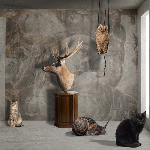
- תערוכה מקורית שלוקחת את הצופה למסע סוריאליסטי לגבולות שבין הידע לדמיון. הזדמנות אחרונה לראות.
- שיר חדש על סיפורו של ד"ר מנחם גורן ואיך הציל מין מהכחדה
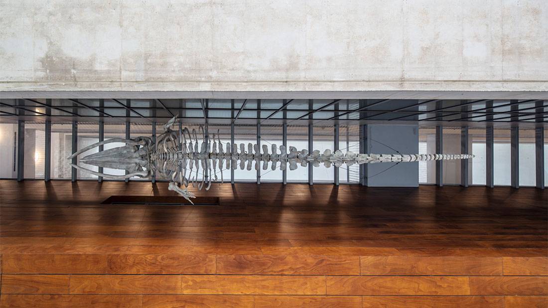
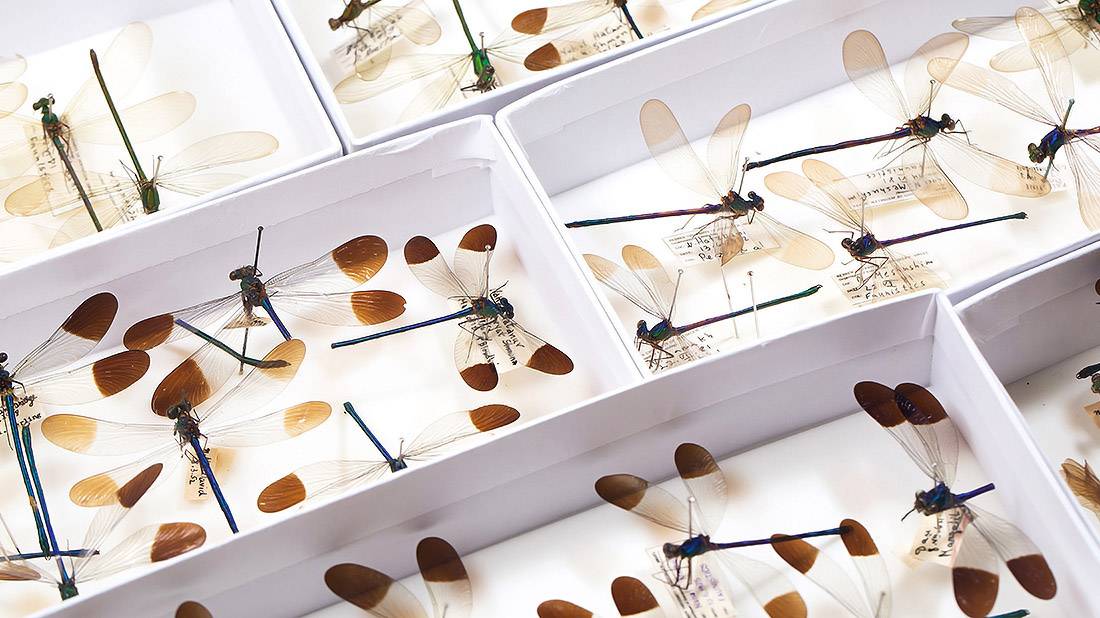
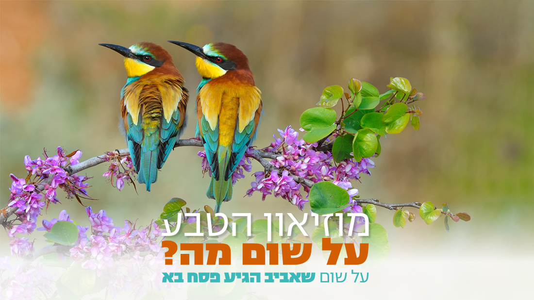
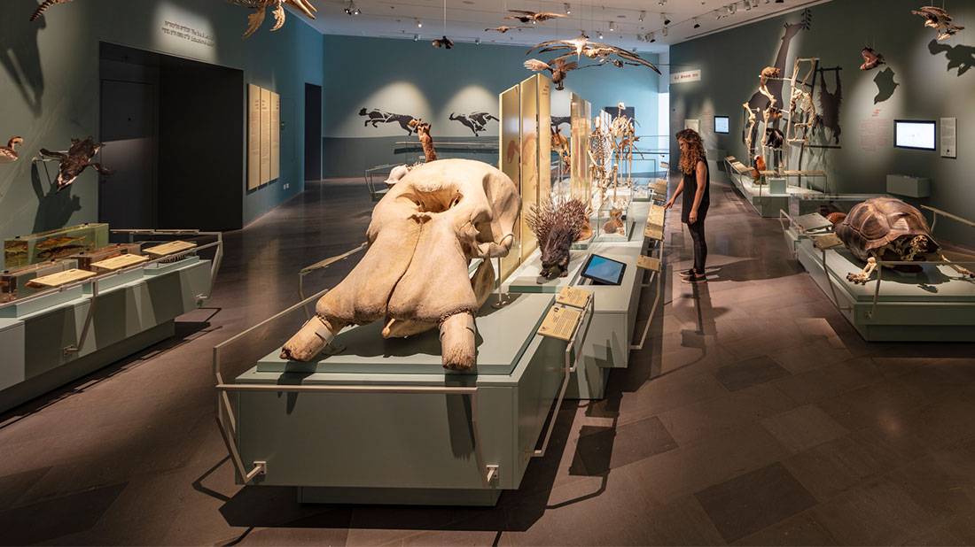
- המוזיאון מציע מיליוני פריטי אוסף המתעדים את עולם החי והצומח של האזור במהלך מאה השנים האחרונות, כמו גם את ההתפתחות וההיסטוריה של המין האנושי.
אירועים

 29-25 באפריל | 10:30; 12:3029-25 באפריל | 10:30; 12:30
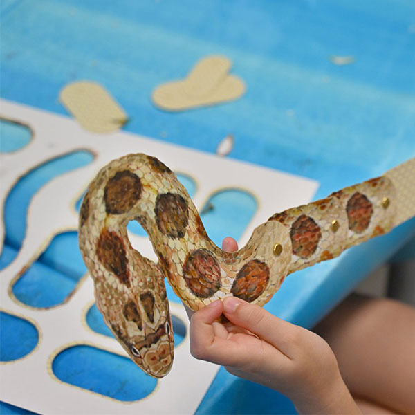
סדנת חקרטבע משפחתית
מדי יום בפסח, בשעות 10:30 וב-12:30 (מלבד שישי) | פעילות למבוגרים וילדים מגיל 6 ומעלה | בשילוב כניסה למוזיאון
כל הפרטים- 29-25 באפריל29-25 באפריל
סיורים מודרכים בגן הבוטני | בעקבות יציאת מצרים
בשעות 9:45 ו-11:30 הכרטיס משולב עם כניסה למוזיאון. בשעה 17:00 ללא כניסה למוזיאון (למעט יום שישי וערב חג).
כל הפרטים - ה׳ | 2 במאי | 19:30ה׳ | 2 במאי | 19:30
נעילת התערוכה HERE BE DRAGONS של נבט יצחק
אירוע לילי בשיתוף "דבק" | דרינק ראשון חינם!
כל הפרטים